Algorithmic Bias in AI Medical Imaging Diagnostic Tools
Models trained on skewed datasets underdiagnose patients from underrepresented groups at scale.
Radiology accounts for 76% of FDA-cleared AI devices, with cardiology trailing at 10% and neurology at 4%. A 2024 European Society of Radiology survey of 572 respondents found nearly half of radiologists now use AI tools day to day, up from roughly one in five just five years back. That's fast growth by any measure, and 97% of these devices reached the market through the 510(k) pathway, a clearance route built for low-to-moderate risk devices. These systems now make judgment calls about who gets flagged for follow-up care, and that mismatch is worth sitting with.
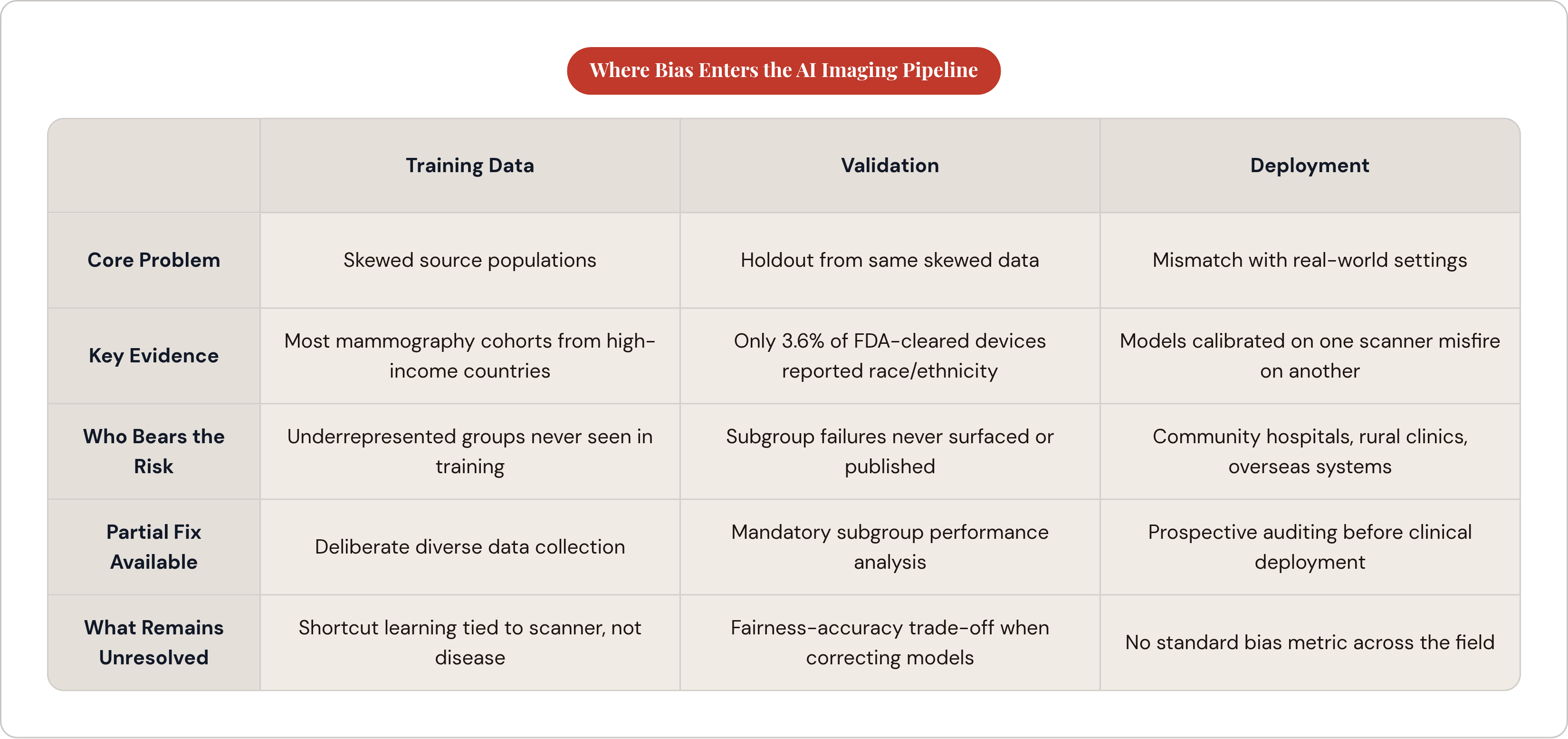
You can't circle one point in red pen and call it the source of the trouble. Bias shows up during data collection and labeling, during training, during validation, during deployment, and even during publication, when studies with clean results get printed and the messier ones quietly don't. Each stage inherits the last one's flaws. A skewed dataset produces a skewed model, that model passes a validation test built on similarly skewed data, and somebody ships it to a hospital its developers never once set foot in.
Deep learning models pick up what researchers call shortcut learning: they learn to tie image features to demographic traits without anyone asking them to. A 2024 study in Nature Communications found models could predict a patient's self-reported race straight from medical images, and part of that signal traced back to acquisition and processing settings, not biology. The model was reading fingerprints left by the machine that took the picture, right alongside whatever diagnostic signal it actually learned to find. That pattern shows up again and again across this pipeline.
Training data decides who gets left out before a single line of code runs. Validation decides what gets caught, and often what doesn't. Deployment drops the finished model into a world that looks nothing like where it was built. Biased outputs in diagnostic imaging touch triage decisions, treatment access, and outcomes, and they land hardest on patients with the least room to push back.
How skewed training data encodes population gaps into models from the start
Models learn from whatever data exists, and existing data reflects who got scanned, where, and by whom. Obvious enough once you say it out loud, but the downstream effects run deeper than the sentence suggests: a 2025 scientometric review in the European Journal of Cancer screened 5,774 mammography AI studies down to 264 that met inclusion criteria, and found most training cohorts came from high-income countries with almost nothing pulled from low-income clinical settings. Somewhere between zero and 25% of those mammography studies even bothered reporting race or ethnicity, and most patients who were identified skewed Caucasian.
Dermatology makes the gap easier to see with your own eyes. HAM10000, the benchmark dataset behind major skin lesion classification contests including the ISIC challenges, underrepresents darker skin tones, and that gap is a documented source of downstream errors. Fitzpatrick17k technically covers all six Fitzpatrick Skin Types but stays lopsided anyway; types four through six sit thin compared to lighter tones. A 2025 study by Joerg and colleagues in the Journal of the European Academy of Dermatology and Venereology looked at 4,000 AI-generated dermatological images and found only 10.2% showed dark skin. Only 15% of the images even accurately showed the condition they were meant to represent, a number worth reading twice.
The gap isn't just who shows up in the dataset; it's also who gets tested in the first place. Documented disparities in who receives routine diagnostic testing mean that some patient populations simply generate less data for models to learn from. A model trained on lab results inherits that testing gap right along with the biology; it learns who the hospital decided was worth testing just as much as it learns actual disease patterns.
A model trained mostly on lighter-skinned patients, wealthy-country hospitals, and patients who got the full workup treats that profile as the baseline. Everyone outside it becomes, statistically, the exception nobody built the system to catch.
What landmark studies found when they tested models across demographic subgroups
Seyyed-Kalantari and colleagues, writing in Nature Medicine in 2021, tested chest X-ray classifiers across MIMIC-CXR, CheXpert, and NIH ChestX-ray14, three of the field's most heavily used datasets. State-of-the-art models consistently and selectively underdiagnosed patients from underserved groups, and the gap widened further for intersectional subgroups, with Hispanic female patients named as one specific example. Underdiagnosis, plainly, means the model looks at a sick patient's scan and calls it fine.
A large pathology study pushed the same question into tissue imaging, using 28,732 whole-slide images from 14,456 patients across twenty cancer types, five medical centers, and three nationwide cohorts. Standard deep learning models showed performance gaps in 29.3% of diagnostic tasks once broken down by self-reported race, gender, and age. Nearly one in three tasks, in other words, carried a measurable gap.
The researchers also found something messier: some of what the models picked up appeared tangled between genuine diagnostic signal and demographic correlation, making it difficult to separate the two. A bigger, more diverse dataset alone won't scrub that out. The signal and the bias are wearing each other's clothes, and nobody's found a clean way to pull them apart yet.
Dermatology gives the starkest real-world stakes. Melanoma incidence runs at 22 per 100,000 among lighter-skinned individuals versus 0.9 per 100,000 among darker-skinned individuals, yet darker-skinned patients face mortality rates 3.75 times higher. That gap already existed before any algorithm touched it. An AI tool trained mostly on light-skin images doesn't invent the disparity, but it can widen it simply by missing the cases it was never shown enough of.
Chest X-rays, pathology slides, and skin images represent three different domains, yet one pattern holds steady across all of them. The populations least represented going in are the same populations where diagnostic errors pile up coming out.
Why validation practices often fail to catch the bias that development introduced
Validation usually runs on a holdout split carved from the same source data used for training. A model trained on mostly Caucasian, high-income-country patients gets tested on more of the same, the top-line number comes back clean, and everybody moves on with their day. That number is answering a much narrower question than anyone in the room realizes, and that gap between what gets asked and what gets answered is where a lot of this trouble hides.
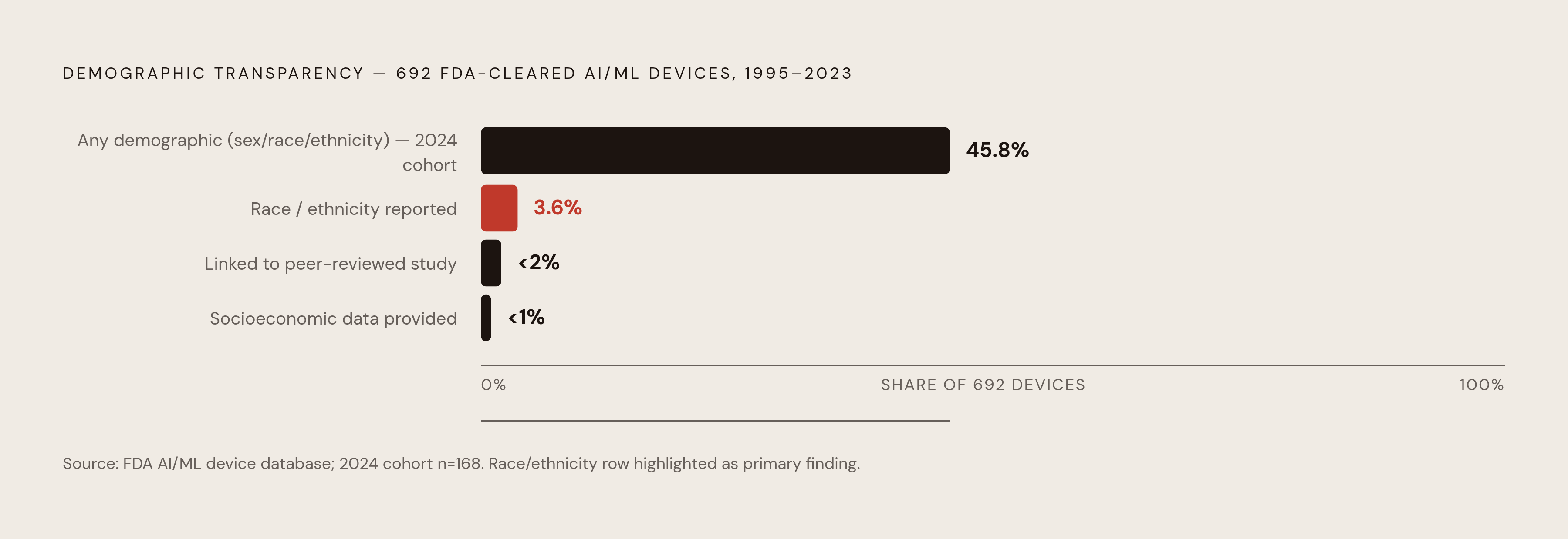
Subgroup performance analysis, breaking results down by race, sex, or age, would catch a lot of this. But it needs demographic labels in the validation data, and those labels are often just missing. A scoping review covering 692 FDA-approved AI and machine learning devices between 1995 and 2023 found only 3.6% reported the race or ethnicity of their validation cohorts. Fewer than 1% gave any socioeconomic information, and fewer than 2% linked back to peer-reviewed performance studies at all. Narrow it to devices authorized in 2024 alone and the number barely moves: only 77 of 168 device summaries, 45.8%, reported demographic breakdowns on sex, race, or ethnicity. That's less than half, for devices cleared this decade, not some relic pulled from a filing cabinet in the '90s.
One might argue the fix is simple: just correct the model for demographic shortcuts and move on. A 2024 Nature Medicine finding complicates that idea considerably. Correcting models to rely less on demographic proxies can reduce how well they generalize to new populations, so the fix trades one failure mode for another instead of patching the original.
Publication bias tightens the knot further. Studies showing strong aggregate performance get published more often than studies showing subgroup failures, so the failures frequently never make it into the literature at all. What does that mean for a clinician or a hospital system evaluating a cleared device? The accuracy number printed on the label may tell you almost nothing about how the tool performs on the actual patients walking through your doors.
How deployment context creates a third source of bias that development never anticipated
Training and validation happen mostly at large academic medical centers, with research budgets, standardized equipment, and patient populations that don't look much like the country at large. Deployment happens everywhere else: community hospitals, rural clinics, health systems overseas, each running different scanners on different patients with different clinical routines.
That gap matters mechanically, not just statistically. Consider again the Nature Communications finding on acquisition parameters carrying demographic signal. A model calibrated on one scanner type or imaging protocol can behave differently the moment it meets another, because part of what it learned was tied to the machine, not the disease it was supposedly diagnosing.
Geography compounds the problem. Algorithms trained mostly on high-income-country data often perform worse in lower-resource settings, and it's usually not biology driving that gap. Clinical context does the driving: the equipment, the imaging protocols, the mix of health conditions walking through the door, none of it represented in training. Mammography AI research grew 311% between 2017 and 2023, climbing from 28 to 115 published studies, yet that growth sits alongside a near-total absence of studies from low-income settings. The literature guiding deployment decisions has a geographic blind spot built right into its foundation.
Shortcut features are especially fragile across this gap. A model that learned to treat a scanner-specific image artifact as a stand-in for disease severity will misfire the moment it runs on different hardware, because the shortcut it leaned on simply isn't there anymore. Human oversight, the safety net everyone assumes is catching these misfires, is often thinner than advertised. Clinicians frequently don't know which populations a given model was validated on, or whether its performance is quietly slipping for the patient sitting in the exam room right in front of them.
What regulators are now requiring and where the gaps remain
Regulators have started catching up, though "started" is carrying most of the weight in that sentence. Regulators have begun issuing guidance aimed at AI-enabled devices across their full lifecycle, with discussions of requiring subgroup performance analysis and demographic-stratified metrics in premarket submissions. New advisory and cross-agency bodies have also taken shape in recent years to help set AI policy principles. The machinery is brand new, which is a polite way of saying it's getting built while the planes are already in the air.
The EU AI Act, in force since August 2024, takes a different tack. Most radiology AI tools qualify as "high-risk AI" under the law, and the core obligations, conformity assessments, technical documentation, mandatory human oversight, apply on a phased timeline. The law puts real weight on deployers, not just the companies writing the software: deployers bear meaningful obligations around compliant procurement, maintaining human oversight, and cooperating with post-market monitoring. None of it is free. Estimated costs run around €29,277 a year per AI unit in operating costs, plus €16,800 to €23,000 per unit just for certification. It's worth asking plainly: can a small rural health system actually absorb that, or does the rule quietly end up favoring whoever already has the biggest compliance department on staff?
And here's the gap nobody's rule closes yet. The 97% of devices cleared through 510(k) before any of this existed are already sitting in hospitals, reading scans, making calls right now. New rules govern what comes next; they don't reach back and re-check what's already running in the wild. Consistent post-market surveillance for demographic subgroup performance and public access to disaggregated performance data remain areas where current requirements leave meaningful gaps.
What actually reduces bias across the pipeline, and what the evidence still can't settle
Diversifying training data is the fix people reach for first, and it's a good one, as long as nobody cheats on the word "diverse." A bigger dataset pulled from the same five hospitals doesn't fix anything; it just makes the same gap heavier. Real diversity takes deliberate work: someone has to go get images from the populations and settings that are currently missing, not just scale up what's already sitting on the server.
Distributed training approaches that keep patient data local while contributing to shared models offer one structural way to widen geographic and demographic coverage, though harmonizing imaging protocols across different sites brings its own complications.
Algorithmic debiasing techniques can reduce measured disparity on whatever demographic axis you're targeting. Consider, though, that Nature Medicine finding on the fairness-accuracy trade-off again: fix bias on one axis and performance can quietly slip somewhere else. Nobody in the field has a clean answer for this yet.
Prospective subgroup auditing before a tool ever touches a patient, treated as a purchasing requirement rather than something developers just self-report, moves accountability to the point where clinical decisions actually get made. Human oversight matters here too, working as an actual safeguard instead of a box to check. A radiologist who knows which populations a model was and wasn't validated on can bring the right amount of skepticism to its output. That requires manufacturers to document validation populations clearly, and right now, that kind of openness stays rare.
So what's still genuinely unresolved? The fairness-accuracy trade-off is one piece, and the lack of any standard bias metric the whole field agrees on is another. Then there's the harder snag buried in the pathology study: how do you handle morphological variation genuinely tied to demographic groups, real biological signal, without either erasing it or letting a model quietly turn it into a shortcut? No clean answer exists yet, and anyone who tells you otherwise is selling something.
Bias in AI medical imaging doesn't get fixed at any one stage, because it was never made at just one stage. Data curation, model design, validation structure, regulatory reporting, and deployment oversight all feed into it, layer on layer. Knowing each piece on its own terms is the minimum anyone would need before they could even start fixing it.